Dear friends, I need help/Feedback!

Assalamoalaikum my dear brother and sisters in medicine. After a long hiatus I want to reinvest sometime in this blog of mine. Life changed, circumstances changes, I changed, everything changed. But this blog stayed the same and that’s not fair in my opinion. So lets start some work. First of all I noticed on my recent visit to a medical book store that they no longer sell MURAD FCPS-1 Mcq books, rather there are some new names in market such as SK Golden, FCPS prep Golden and what not. In today’s digital world copyright is a huge issue. Although all these books print MCQs recalled by past test takers, I still can’t copy the questions and paste them here in my blog despite the fact that I write my own original explanations. I tried to look for Facebook groups where I can find past papers without any copyright issue but I wasn’t very successful. So the running on this blog now depends on two things.

  1. Reader provided MCQs or
  2. Writing short explanations without the questions (this would cover a small topic that appeared in the past papers starting from the most recent July 2021

So I will start with the short explanations without posting the questions and also wait to get more input from readers. Lets see how it works out.

PS: It would be nice if some of you can guide me to some free resources where I can get some input of past papers.

Leave a comment

Filed under Uncategorized

HIV or AIDS? What’s the difference?

Often used interchangeably in layman language, HIV positivity and AIDS are not the same thing and that’s why the examiners want to know: are you a doctor or a layman?

So first things first. HIV is Human Immunodeficiency Virus and AIDS is Acquired Immunodeficiency Syndrome. Only an HIV-positive patient can develop AIDS but NOT all HIV-positive patients have AIDS. HIV-positivity doesn’t automatically mean that a patient has AIDS. Only when the HIV- virus attacks and weakens the immune system of an individual, he/she develops AIDS. There is a very specific criteria to diagnose a patient with AIDS while a simple PCR laboratory test can label a patient as HIV positive. Since patients with AIDS are Immunodeficient so they can easily get infected by organisms that otherwise do not cause infections in an immunocompetent person. So if a question states that a patient is HIV-positive but DOES NOT have AIDS then consider him/her as an average immunocompetent person.

Now we come to our question which is if a woman who is HIV positive but doesn’t have AIDS develops otitis media, what would be most likely organism? Since we already clarified that a person who doesn’t have AIDS, is not affected by opportunistic organisms. So please don’t jump to the conclusion that since she is HIV positive she will be affected by all the strange named opportunistic organisms. So we use our common sense here. In this case just recall the three most common organisms that cause otitis media in immunocompetent persons:

Streptococcus pneumoniae (most common)

Haemophilus influenzae

Moraxella (Branhamella) catarrhalis.

These three organisms are responsible for more than 95% of all Acute otitis media cases with a bacterial etiology.

Leave a comment

Filed under FCPS-1 Concepts., Uncategorized

Question 12 (Nerve & Muscle)

Question: The blockage of voltage sensitive sodium (Na) channels can be brought up by

a. Tetradotoxin

b. Opium

c. Curare

d. Parathion

e. Hemicholinium

Answer: a. Tetradotoxin

Explanation:

Tetradotoxin is a potent neurotoxin. It is found in certain fish and is a sodium channel blocker. It binds to voltage-gated Na channels in the nerve cell membranes and inhibits the action potentials form propagating and so prevents signal travel from cell to cell resulting in paralysis.

Curare is a muscle relaxant that blocks the nicotinic acetylcholine receptor (nAChR).

Opium is a narcotic drug that acts of opioid receptors

Parathion is an organophosphate insecticide that acts by inhibiting acetylcholinesterase.

Hemicholinium interferes with the synthesis of acetylcholinesterase by blocking reuptake of choline at the synaptic cleft which is required for the synthesis of acetylcholinesterase.

Leave a comment

Filed under Uncategorized

Question 11 (Cell Physiology)

Question: Regarding cells all of the following are true except

a. intermediate filaments are one of the component of cytoskeleton

b. Mitochondria is self-replicative

c. Cells involved in protein synthesis have well developed smooth endoplasmic reticulum

d. Lysosomes engulf worn-out competes of the cell


Answer:  c

Explanation:

Its worth remembering functions of the different components of the cells.

Endoplasmic reticulum is the “transport system” of the cell and is essentially of two types. Rough endoplasmic reticulum (RER) and Smooth endoplasmic reticulum (SER). The RER is rough because it has ribosomes attached on its surface giving it a rough surface. Function of these ribosomes is to produce protein. Since the SER has no ribosome attached to it, so it cannot produce protein. Only RER can.

Leave a comment

Filed under Uncategorized

Question 10 (Neuroanatomy)

Question: Inability to perform alternating movements of the hands is referred to as:

a. Dysdiadochokinesia

b. Dyskinesia

c. Romberg’s sign

c. Chorea

e. Athetosis


Answer: a

Explanation:

Dysdiadochokinesia is the inability to perform alternating movements of the hands. With dysdiadochokinesia think cerebellum. Dysdiadochokinesia can occur in any condition that affects the cerebellum. In other words dysdiadochokinesia is a cerebellar sign. During neurological examination if you want to check out the cerebellar function of the patient, one of the tests is to ask the patient to place one hand over the other and have them flip one hand back and forth as fast as possible. Patient with cerebellar dysfunction can’t usually do that.

Watch a quick video here on YouTube if you are interested.

Dyskinesia refers to involuntary movements that a person cannot control

Romberg’s test checks if he patient can stand steadily with his eyes closed or not. The patient is asked to stand with their feet close together and then they are asked to  close their eyes. (Make sure to stand near by in case patient falls upon closing the eyes). If the patient can stand with eyes open but sways on closing the eyes, it means Romberg’s sign is positive.  Romberg’s sign is positive in sensory ataxia and negative in cerebellar ataxia. That is the patient is bound to sway/fall with eyes open or close if he has problem in his central part of his cerebellum. So Romerg’s is said to be negative. But if the problem lies in sensory tracts (proprioception) or there is a vestibular problem, the patient might be able to stand steady with the eyes open (visual input helps the patient to maintain steady stance), but sways or falls on closing the eyes (lack of visual input).

Chorea is an abnormal involuntary movement  derived from the Greek word “dance”. It is characterized by brief, abrupt, irregular, unpredictable, non-stereotyped movements.

Athetosis means slow, purposeless, and involuntary movements of the hands, feet, face, tongue, and neck (as well as other muscle groups).

Leave a comment

Filed under Uncategorized

Question 9 (Head & Neck)

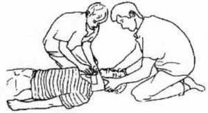
Question: You are a qualified trauma surgeon traveling in your car on a highway having your  well-equipped kit with you. At the scene of accident you find an unconscious driver. After clearing his airway your next step of management would be?

a. Have an IV line

b. Do tracheostomy

c. Pass endotracheal tube

d. Secure cervical spine

e. Manage shock

Answer: d

Explanation: 

Lets dissect the answers one by one.

Have an IV line: there is no use having an IV line right there at the road side. You might say that the first things are always Airway  (which this surgeon, that is you) have cleared, Breathing, Circulation. Now you might argue that passing an IV line is included in Circulation. There are two things there. First, there is no mention of a cardiac arrest (which can compromise circulation), second even if the patient is in cardiac arrest  (which he is not), then your priority should be chest compressions to maintain the circulation and not an IV line.

Do tracheostomy: Haven’t you been clearly told that he has cleared the airway? A tracheostomy is required only if the upper air way is blocked and cannot be cleared. So there is no need of tracheostomy in this situation

Pass endotracheal tube: Since the airway is clear and there is no mention about the patient NOT BREATHING (which requires ventilation) so there is not need to pass endotracheal tube.

Manage shock: Shock?? Where is shock in this question? No where. So if something is not mentioned, don’t bother cooking it up. (Although some FCPS-1 questions are absurd enough that to answer then you have to cook the missing bits of questions (a problem with CPSP) up and then answer it)

Secure cervical spine: Ah…here comes our correct answer. Now whenever you find an unconscious trauma patient (like in this question), after clearing off the airway and making sure he is breathing and not dead, stabilise his cervical spine and DO NOT manipulate the neck of the patient in any case. Even when securing the airway you have to very careful about cervical spine. Because if you don’t secure the cervical spine first, instead of saving the patient’s life, you might actually kill him or get him quadriplegic for life if the cervical vertebrae were damaged in the accident and you put further insult to injury and break his cervical spine. So secure it first!

In short the management might include  all of the above options, but the emphasis is about priorities, that is which step should be done first.

Securing the spine

Securing the spine

Leave a comment

Filed under Anatomy, Head & Neck

Question 8 (Abdomen)

Question: A patient comes to you with portal hypertension and dilated abdominal veins, which vessel is involved?

a. Hepatic Vein

b. Inferior vena cava

c. Portal vein

d. Hepatic artery

e. Superior mesenteric artery

Answer: c

Explanation:

This is the kind of MCQ in which the answer lies in the question, look at the phrase “portal hypertension” in the question, well the best guess (that is if you are clueless about what portal hypertension actually is!) would be portal vein even without knowing the anatomy. But since we are here to become good doctors so its better to leave the wild guessing game aside and learn some basic things about the portal anatomy.

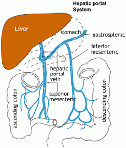
The portal system The portal system

In simplest of words, the portal vein receives all the blood from the gut and takes it to the liver where nutrients are processed and blood detoxified by the liver and the blood is then dumped into the inferior vena cave which takes it to the heart. Since the blood from the gut does not directly go to the heart (instead goes first to another organ, i.e, liver), it is therefore called the portal (carrier) system.

The portal vein blood gets into the liver sinusoids and from here the hepatic vein drains it  and enters the inferior vena cava. This the normal pathway. If the liver sinusoids are distorted like in cirrhosis of liver, the blood from portal vein has trouble passing through the liver. Now the pressure in the portal vein rises as the forward resistance is high due to distorted sinusoids. This is the portal hypertension.

The blood from the gut however still has to get back to the heart no matter what. So the collaterals between gut veins and other veins which drain directly into the inferior vena cava, open up. This way most of the blood from gut can bypass the portal system/liver and reach the heart directly. This results in the collaterals to enlarge. Three important areas where this shunting of blood from portal system to systemic veins (bypassing the liver) occurs are:

At the lower end of oesophagus (causing esophageal varices)

At the rectum

Over the anterior abdominal wall (like in this question, where abdominal walls are dilated)

Leave a comment

Filed under Abdomen, Anatomy

Question 7 (Thorax)

Question: During exhaustion, a person uses the accessory muscles of inspiration. The muscle which primarily increases the anteroposterior diameter of thoracic cage is?

a. Sternocleidomastoid

b. Scalene Anterior

c. Internal Intercostal

d. External Intercostal

e. Diaphragm


Answer: d

Explanation:

Its important to know about muscles of respiration as questions often come up in exam about them.

During quiet breathing, the predominant muscle of respiration is the diaphragm. Expiration during quiet breathing is predominantly a passive phenomenon, as the respiratory muscles are relaxed and the elastic lung and chest wall return passively to their resting volume, the functional residual capacity.

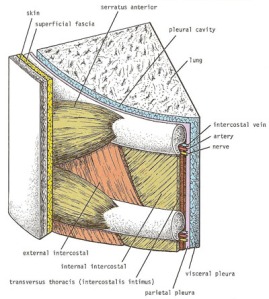
However, during exercise or exhaustion like in this question, many other muscles become important to respiration. During inspiration, the external intercostals raise the lower ribs up and out, increasing the lateral and anteroposterior dimensions of the thorax. The scalene muscles and sternomastoids also become involved, serving to raise and push out the upper ribs and the sternum.

During active expiration, the most important muscles are those of the abdominal wall (including the rectus abdominus, internal and external obliques, and transversus abdominus), which drive intra-abdominal pressure up when they contract, and thus push up the diaphragm.

The internal intercostals assist with active expiration by pulling the ribs down and in, thus decreasing thoracic volume


intercostal_muscles

Leave a comment

Filed under Thorax

Question 6 (Lower Limb)

Question: A mass along the lateral wall of the pelvis results in the paralysis of the medial muscles of the thigh. Involvement of which of the following structures is most likely cause of this paralysis?

a. Exteranl iliac artery

b. Femoral Nerve

c. Obturator Nerve

d. Sciatic Nerve

e. Superior gluteal nerve

Answer: c

Explanation:

The key to solve any MCQ is to reach the root question quickly. In this question the plain simple thing being asked is about the innervation of medial thigh muscles, in a twisted way though!

So lets jump to the answer now. First, which are the medial muscles? The adductors obviously cause they lie medially so they can pull the thigh medially (adduct it). Next which nerve supplies the adductor of hip (or medial muscles of thigh)? Its the obturator nerve. From the question we can see that there is mention of a mass running along the lateral pelvic wall. Obturator nerve runs along the lateral wall of the lesser pelvis to enter the thigh later. So a mass along lateral wall of pelvis can damage the obturator nerve.

Can you name the medial thigh muscles (the adductors)?

These are:

Adductor brevis

Adductor longus

Adductor minimus

Adductor brevis

Pectinues

Gracilis

cg0LzJ_uE6w8m08eluTsqQ_m

Obturator Nerve


Leave a comment

Filed under Anatomy, Lower Limb

Question 5 (Upper Limb)

Question: A person can’t abduct his arm after fall on the out-streched hand; however he is able to abduct it after the initial 90 degrees of passive abduction of his arm. The most likely muscle involved is?

a. Trapezius

b. Deltoid

c. Pectoralis major

d. Supraspinatus

e. Teres Major

Answer: b

Explanation:

Remember that in  normal subjects the supraspinatus initiates the first 15 degrees of abduction along the vertical plane. The deltoid functions from 15 to 90 degrees, while synergistic actions of the trapezius and serratus anterior abduct from 90 to 180 degrees by rotating the scapula laterally.

So our patient in this question is unable to abduct this arm till 90 degrees only (indicating a paralysed deltoid), and then from 90 degrees onwards his trapezius and serratus anterior muscles take over and perform the rest of abduction.

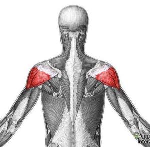
A little bit of more information about the Deltoid which is a favourite muscle in medical exams:

Deltoid is supplied by the Axillary Nerve (C5, C6)

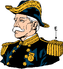
I like to think of deltoid as the epaulet (the shoulder ornament you see on soldier uniforms, picture follows).

The deltoid muscles.

The deltoid muscles.

epaulet

The epaulet that overlie the deltoids.

Leave a comment

Filed under Anatomy, Upper Limb